ULTRASONIC – TRIPLEX
Heart triplex
The Heart Triplex (heart ultrasound) is an imaging, non-invasive examination of the heart. With the help of ultrasound, we have the ability to clearly visualize the structures of the heart, such as the cavities, valves, blood flow, pericardium, walls and their movement as well as to determine their integrity and functionality. The examination is painless, is carried out by the doctor and is done using the most modern, latest generation equipment, having the possibility of applying, not only the traditional techniques (2D resolution, M-Mode, Doppler, Triplex) but also all new advanced techniques such as dynamic echocardiography (stress echo), contrast echocardiography (contrast echo), the tissue doppler imaging, 3D analysis and deformation analysis, with the strain rate and the speckle tracking, tools, which, in conjunction with our doctor’s expertise in echocardiography, place our center among the highest quality cardiac ultrasound laboratories in the country.
Aortic triplex
The triplex aorta it is also an imaging, non-invasive examination, with which the morphology of the lumen and the flow of blood within the aortic vessel are checked and the existence of stenosis of the vascular lumen and the changes that the blood flow undergoes due to this stenosis and also the presence of dilatation and soft spots of the vessel, as in aneurysms. It is also possible, with the use of appropriate techniques, to study the hardness of the vessel and thus assess the existence of atherosclerotic vascular disease.
Contrast echo
The use of contrast agents in echocardiography (contrast echo) can identify possible hidden communications between the heart chambers and better highlight the motion of the heart walls, in patients who are difficult to assess due to their anatomy. Their use has applications in the investigation of congenital heart anomalies but also in the diagnosis of coronary artery disease in difficult cases. In the Stress Echo, with the use of special software available in our clinic, is also possible, with the use of very low frequency ultrasound (Low MI), to clearly demonstrate the perfusion of the walls and the existence of Coronary artery disease with great reliability, in difficult cases where this is not obvious. The use of contrast agents requires special certification and sophisticated software, which our practice has.
Assessment of carotid arteries
Along with the ultrasound examination of the heart, there is the possibility of assessing the carotid arteries, for the possible presence of stenosis of the lumen and blood flow disorders. This assessment does not replace the Carotid Triplex, which is a radiological procedure and is carried out by a specialist radiologist with vascular training.
EXERCISE TESTS – CARDIAC ISCHEMIA CONTROL
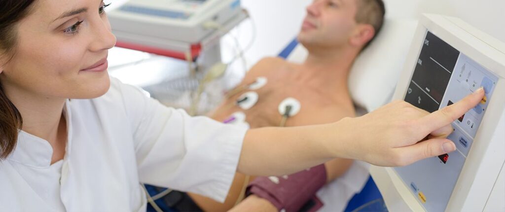
Fatigue test
It is a simple, painless and bloodless test, which contributes to the diagnosis and monitoring of coronary artery disease. The two main features of the fatigue test are the performance of controlled physical exercise by the subject on a treadmill and continuous electrocardiographic monitoring on a screen both during and after exercise. It helps the doctor to assess the subsequent functional disorders of the narrowing of the lumen of the coronary arteries.
Stress – contrast Echo
Dynamic echocardiography belongs to the newest heart ultrasound techniques and is mainly aimed at the early and effective diagnosis of coronary heart disease. With dynamic ultrasound instead of electrocardiographic changes we try to identify the mobility deficits. In other words, we monitor the movement of the walls of the heart during special stress with an increase in the frequency of heart beats, as if we were exercising. How is this done? By administering a small amount of a special drug, we make the heart work faster so that we create a small stress on the heart and increase its need for oxygen, i.e. more blood is required to bleed. If there is a stenosis in an artery and the blood does not flow well from there, then there will be a problem in the movement of this part of the heart, which we will see in the ultrasound, as a result of which we will diagnose Coronary heart disease early, without waiting or having the doubt whether or not the ECG of the examinee will change. With the additional use of contrast agents (contrast echo) the sensitivity of the method increases and we can see the perfusion of the walls better.
It has therefore been found that with this method we can safely diagnose up to about 90% of patients with Coronary Artery Disease in contrast to a common fatigue which is 67%.
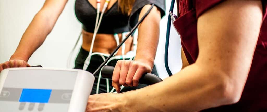
Exercise Echo
Dynamic exercise echocardiography (or Exercise Echo) is an important test for demonstrating myocardial ischemia and documenting the existence of coronary artery disease in patients with no previous history. The basic principle of the examination is the same as the stress echo, but without, intravenous drug use. That is, it is based on monitoring the movement of the walls during exercise, which is achieved by using a treadmill or bicycle. Physical exercise, the results of which are enhanced when combined with handgrip (mild isometric exercise after the end of aerobic exercise), is an important factor in increasing the sensitivity of the method, as long as the study of the walls can be assessed with powerful analysis software tools, provided only by modern ultrasounds of the latest generation. Our clinic has such equipment and is able to produce very reliable results, having gained vast experience from carrying out over 3000 tests of this kind.
HOLTER – MONITORING DEVICES
Holter Rhythm
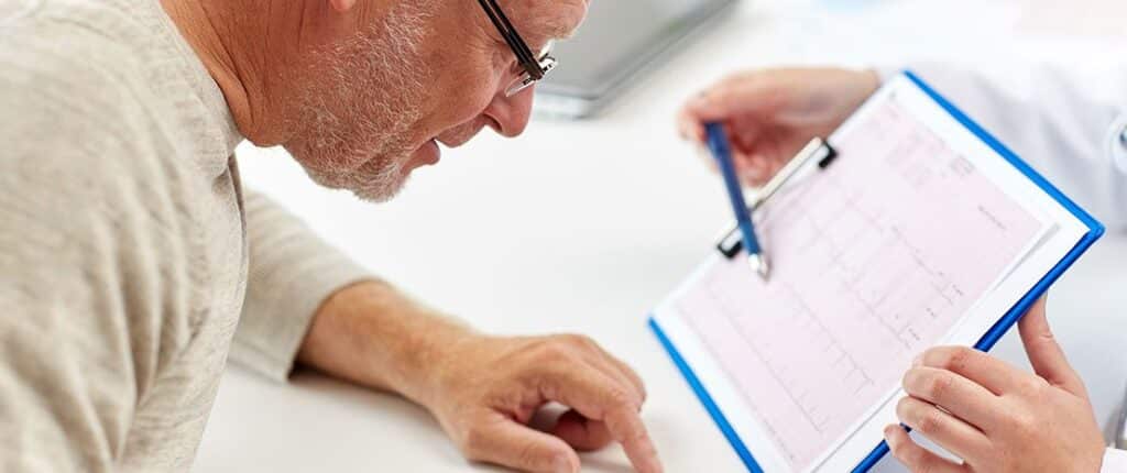
The rhythm Holter is a continuous 24-hour electrocardiogram recording of the examinee, thus increasing the chances of finding possible transient disturbances of the heart rhythm, which could not be investigated with the simple electrocardiogram, since this is a short recording of only a few seconds.
It is a basic examination in the diagnostic approach to arrhythmias and disorders of the excitatory system of the heart. Holter Rhythm also plays an important role in investigating symptomatology related to palpitations, tachycardia, prefainous, syncopal, or syncopal episodes. With the same examination, there is also the possibility of detecting silent ischemia, i.e. myocardial ischemia that is not noticed by the patient.
The test is non-invasive and completely painless and is carried out by placing a few adhesive electrodes on the examinee’s chest and a small, light, portable and very discreet device on his belt or around his neck. It remains in place for 24 hours, during which the examinee carries out his normal daily activities. The device is then returned to be analyzed by the physician.
In case this is requested by the attending physician, the recording of the heart rate can last up to 48 hours.
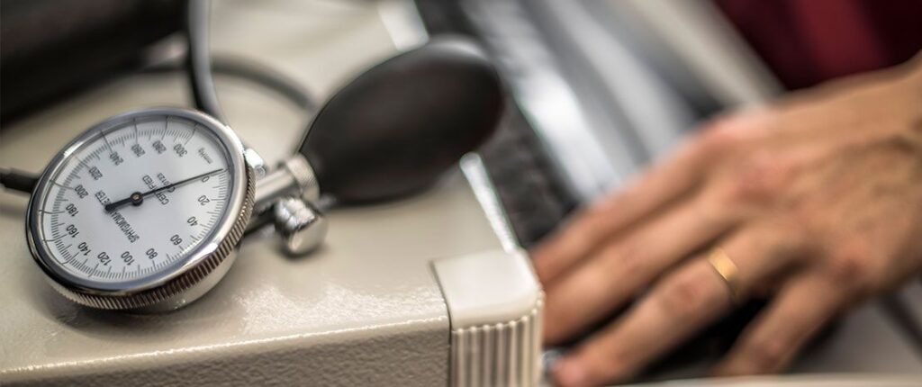
Holter Pressure
The blood pressure holter is a portable electronic blood pressure monitor that automatically measures blood pressure every 15-30 minutes for 24 hours. It consists of a cuff that wraps around the arm and a cell phone-sized device that connects to the cuff and is worn on the belt or slung over the shoulder. The application of this technique provides the possibility of multiple pressure measurements away from the “stressful” environment of the clinic. This gives a complete picture of the pressure in the usual conditions of a working day, during a day’s work, at home and while sleeping.
Sleep Holter (Portable Nocturnal Oximetry)
This is a small portable oximetry device, provided to the examinee, in case of suspected sleep apnea syndrome. The device can be easily placed before going to bed while not burdening the examinee, since it is worn on the wrist with a fabric wristband. After the device is returned, special software analyzes the patient’s oximetry and heart rate during sleep, reaching useful conclusions about the possibility of sleep apnea. This device, by itself, cannot establish a diagnosis of sleep apnea syndrome, and in case of suspicion, a polysomatic sleep study will then be required.
Portable sleep study system
A sleep study, on the other hand, is a more comprehensive examination method, which is used to diagnose sleep apnea and other sleep disorders. The patient, in collaboration with us, wears the device at home, during the night, to record and then study his respiratory function while he sleeps. This method is a small polysomatic sleep recording study, which is the reference test in the diagnosis of sleep apnea, while the evaluation of the results is done in collaboration with specialized pulmonologists. Symptoms that may lead to a sleep check are snoring, daytime sleepiness, morning headaches, poor memory, lack of concentration, sexual problems and insomnia.
multi– Holter
The SOMNOtouch system is a 24-hour simultaneous blood pressure, rate, oximetry, sleep apnea monitoring sensor and arterial stiffness index (PWV) device. It is small in volume and weight with high comfort for the patient and minimizes the differences caused by changes in body position. This device is the only one that can measure blood pressure, without the use of a cuff, while at the same time it gives us live data for the assessment of arterial stiffness and atherosclerotic vascular disease. The great thing about this device is that it can simultaneously combine several recordings into one, without subjecting the examinee to the stress of continuous inflation of the cuff while giving the examiner the possibility of drawing many useful conclusions.
DIAGNOSTIC CONSUMPTION TESTS

In our practice, a test can be performed to investigate the causes of syncope (brief loss of consciousness of a few seconds), presyncope, fainting episodes and diseases of the electrical conduction system of the heart. The cardiological assessment of these patients requires a series of tests, including:
- Carotid massage (test, under continuous electrocardiographic and hemodynamic monitoring, to investigate hypersensitive carotid sinus syndrome )
- Δοκιμασία ορθοστατισμού (δοκιμασία, υπό συνεχή αιμοδυναμική παρακολούθηση, για τη διερεύνηση ορθοστατικού συνδρόμου και ορθοστατικής ταχυκαρδίας)
- Atropine test (administration of atropine to investigate the presence of sinus disease )
- Leaning test (Tilt test) (reclining bed test, under continuous electrocardiographic and hemodynamic monitoring, to investigate syncope and malignant vagotonia ).
All of the above tests are available in our practice (except the recline test, which will be available soon).
SPECIAL BIOMETERS
Hammer index
The Hammer (or Tibial) index ( ABI) is an assessment indicator of the existence of peripheral vasculopathy, manifested by the occlusion of the arteries of the lower extremities. The measurement of the index, which requires the measurement of the systolic pressure in the two lower extremities and the right upper extremity, is easily done in our clinic using the device ABPI MD (MESI), the first automatic device that performs its measurement Hammer index, based on oscillometry method. In this way, the diagnosis of peripheral arterial disease can be made within just 1 minute.
Body composition analysis
By the term body composition analysis, we mean the analysis of body weight in relation to body fat, other tissues and the amount of body water. To calculate the composition and body type, we use special biomagnetic body meters, in order to determine whether the body of each person is in good physical condition or whether there is an issue of metabolism or obesity. The analysis of body composition can also give us useful conclusions for the adequate hydration of the examinee and set the goals of medical and dietary interventions.
Basic metabolism
Basic metabolism is the calculation of the minimum energy that our body needs to perform its basic functions. Its measurement used to require very expensive machines and prolonged measurements in a closed environment. The measurement of the basic metabolism can now be done in our clinic, very simply, by breathing and using the innovative Breezing device. This device is an “intelligent” and very easy to use indirect calorimetry meter, which gives us for the first time the possibility to have in our hands directly and accurately, the real information about the metabolism. Its calculation is very important for the assessment, diagnosis and treatment of complex metabolic problems, which accompany several morbid conditions.
Physical activity meters
Fitness trackers are small wristwatches that automatically measure your activities during the day and night. Heart rate, sleep duration, calories burned, steps taken and distance traveled daily can be automatically recorded and studied online. So the doctor can immediately assess the level of physical activity you have and give you useful advice for a proper healthy lifestyle according to the needs of your body and the health goals, defined by the guidelines of scientific societies.